Musculoskeletal Imaging
Our research emphasis in musculoskeletal (MSK) imaging is on advanced imaging of degenerative musculoskeletal diseases, particularly those with a large burden for patients and society such as osteoporosis, but also on improved visualization of traumatic musculoskeletal injuries.
Sensitive and accurate imaging algorithms are the key for early diagnosis and initiation of appropriate therapy. Therefore, our goal is to develop, improve and validate novel imaging methods, imaging protocols and methods for image analysis, and to apply them in clinical studies. Many of our imaging techniques are aimed at visualizing and quantitatively measuring tissue composition changes and pathological tissue processes in musculoskeletal disease.
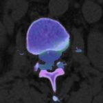 One of our key goals is to develop phantomless dual-energy computed tomography (DECT)-derived quantitative bone mineral density (BMD) assessment which can be performed in routine imaging so that BMD may be measured in every scan to improve diagnosis of osteoporosis.
We also develop DECT algorithms to visualize trabecular BMD distribution for improved presurgical assessment, color-coded visualization of bone marrow edema ("bone bruise") following trauma and color-coded visualization of anatomical structures such as intervertebral discs using. To improve and validate DECT-based reconstruction algorithms for these purposes, we utilize information from magnetic resonance imaging (MRI) and other imaging approaches.
Vascular Imaging
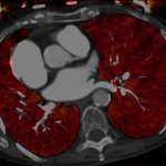 Vascular computed tomography (CT) is becoming an increasingly important diagnostic tool with growing clinical utility. Our research aims at translating state-of-the art dual-energy CT techniques into clinical practice and improving image quality. The implementation of the noise-optimized virtual monoenergetic imaging (VMI+) technique revealed several advantages for vascular imaging due to enhanced contrast conditions. In this context, our research demonstrated the additional value of VMI+ for CT angiography of the pulmonary arteries, active arterial bleeding of the abdomen, and endoleak detection.