The cruciate ligaments are major stabilizers of the knee joint and frequently injured, especially among athletes. In this context, early reconstruction has been shown to reduce the risk of further knee damage and accelerate the return to physical activity. MRI is currently the gold standard for diagnosing cruciate ligament injuries, but its availability is limited, and certain patient conditions may contraindicate its use.
Advances in dual-energy CT technology now offer superior spatial resolution and material differentiation capabilities, which allows for the depiction of soft-tissue structures. In a recently published study by Leon Gruenewald, MD, we evaluated novel postprocessing algorithm designed for visualizing collagen structures for its capabilities in assessing the cruciate ligaments. We included 85 patients who had undergone non-contrast third-generation dual-source dual-energy CT followed by supplementary MRI and/or arthroscopic inspection of the knee joint within 14 days in this retrospective study. All CT images were acquired on a third-generation dual-source CT system in dual-energy mode. Following image acquisition, color-coded collagen reconstructions were created on a Siemens syngo.via VB50 using an experimental algorithm. Independent evaluation of the images was performed by five radiologists blinded to all clinical information. Color-coded collagen reconstructions showed high overall sensitivity (90%), specificity (78%) and accuracy (81%) for the identification of injury to the cruciate ligaments compared to MRI or arthroscopic inspection as the gold standard, indicating that dual-energy CT may serve as a readily available screening approach for patients with acute trauma to the knee when injury to the cruciate ligaments is suspected.
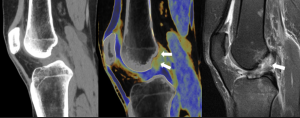
Dual-energy CT colored collagen map shows lack of the blue colored collagen signal in the proximal portion of the ACL in case of an ACL tear (arrows), which was confirmed by additionally performed MRI.